Gel Documentation

Gel documentation systems, commonly referred to as 'gel docs' or 'gel imagers,' are indispensable tools used in molecular biology labs for recording and analyzing the outcomes of gel electrophoresis and membrane blotting experiments. These systems are essential for visualizing stained or labeled nucleic acids and proteins within media like agarose, acrylamide, or cellulose. Depending on the specific application, throughput requirements, and sample type, gel documentation systems are available in various configurations.
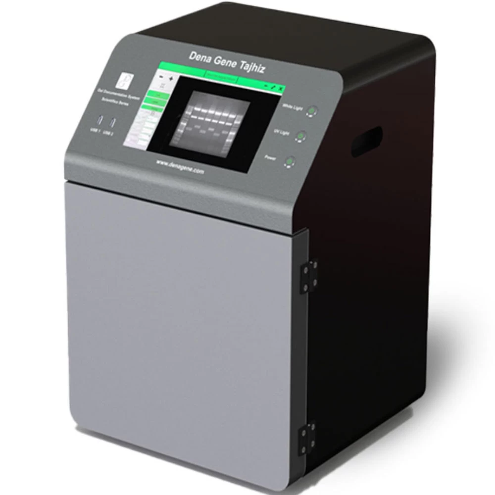
A gel documentation system is primarily utilized in molecular biology labs with the objective of imaging and documenting nucleic acids and proteins suspended in agarose or polyacrylamide gels stained with fluorophores such as SYBR green or ethidium bromide. It is also known by other names like gel imager, gel imaging system, and gel doc system.
Gel documentation systems play a pivotal role in molecular biology research, equipping researchers with the necessary tools for accurate and efficient visualization, documentation, and analysis of nucleic acids and proteins. Molecular biology and biochemistry laboratories heavily rely on gel documentation systems as vital instruments for viewing and recording protein and nucleic acid samples that have undergone separation through gel electrophoresis.
Gel documentation systems enable researchers to visually observe the bands of DNA, RNA, or proteins within a gel, providing a clear representation of the outcomes of gel electrophoresis.
Documentation and Record Keeping
This technology simplifies the process of collecting and storing high-resolution photographs or data of gels, ensuring reliable documentation and record-keeping of experimental results.
Gel documentation systems often include software that allows for quantitative analysis of bands, enabling researchers to determine the size and intensity of DNA, RNA, or protein bands accurately.
By digitally capturing gel images, researchers can achieve more precise and consistent results. Digital data is easier to manage, share, and analyze compared to traditional film-based approaches, enhancing data accuracy and reproducibility.
Digital photos captured by gel documentation systems can be conveniently stored for future reference, facilitating the retrieval and comparison of results over time. This is particularly valuable for long-term research projects or when replicating studies.
Certain gel documentation systems offer the capability to view gels at different wavelengths, which proves useful for various stains or fluorescent markers employed in the experiment.
Gel documentation technologies eliminate the need for time-consuming processes like film development. Researchers can quickly capture images, expediting the entire experimental procedure.
Many gel documentation systems are equipped with UV transilluminators for fluorescence imaging, allowing the detection of fluorophores such as ethidium bromide or SYBR Green, commonly used for nucleic acid staining.
The seamless exchange of digital photographs among researchers and collaborators, as well as their integration into papers and presentations, fosters effective communication within the scientific community.
Gel documentation systems are highly adaptable and can be utilized for a wide range of tasks, including protein, RNA, and DNA electrophoresis, as well as Northern, Southern, and Western blotting.
The instant visibility of gel data provided by gel documentation systems enables researchers to conduct quality control checks during or immediately after the electrophoresis process, minimizing the likelihood of errors or the need for repeated tests.
Different configurations and specifications are available in the gel doc system, depending on the sample type and throughput. The gel doc system consists of various systems, including the chemiluminescence imaging system, digital gel imaging systems, automated blot analysis, and multiplex fluorescence imaging systems.
In the life science laboratory, western blotting is a crucial technique used for protein separation based on molecular weight. The chemiluminescence imaging system is employed as a detection method for western blotting due to its high sensitivity. Imaging systems utilizing this technique enhance speed, sensitivity, and signal stability.
Digital gel imaging systems are utilized to capture and measure stained acrylamide and agarose gels using a digital platform. These systems not only offer efficient data storage but also enable accurate quantification of samples. They employ different types of detectors, such as visible light, Ethidium bromide (UV), fluorescence, chemiluminescence, and densitometric detectors, for quantitative analysis of protein and nucleic acid bands, microplates, and dot blots. This system simplifies the image acquisition process by providing options for auto-exposure and auto-focusing.
Automated blot analysis systems are sensitive imagers that utilize specialized software for analyzing western and nucleic acid blot data. These systems automate tasks such as focusing, light selection, exposure, and acquisition. They offer various detection options, including UV, infrared, RGB color, fluorescence, and chemiluminescence. With multiple fluorescent channels, these systems can measure multiple targets simultaneously.
Multiplex fluorescence imaging systems are used to illuminate blotting membranes and detect signals above background noise. They capture the blot image and analyze the signals. Most systems consist of a light source, emission filters, and a photosensor (CCD or CMOS camera). Light sources such as RGB, UV, and white light are commonly used. Certain systems can detect and image multiple fluorescent signals simultaneously through multiplexing. Some fluorescence imaging systems can also detect radioisotopic, luminescent, and colorimetric signals. Confocal optics may be employed in some systems to enhance sensitivity when detecting targets present in low concentration.
